For a full-arch implant restoration, the impression has one main job: to transfer the three-dimensional position of every implant from the mouth to the laboratory, so that the definitive framework fits passively. A small transfer error that a single crown might tolerate can add up across a rigid multi-unit framework. There are two broad ways to record implant positions: a conventional (analog) impression with impression copings and an elastomeric material, or a digital impression in implant dentistry, made with an intraoral scanner and scan bodies. This article explains how each workflow works, where errors arise and how to verify the result, for dentists and dental students.
Why full-arch implant impressions are demanding
Several features of the edentulous arch make the transfer step demanding:
- Several implants, often at different angles. Angled implants and deep platforms make coping removal and seating more difficult.
- A long span. Small errors at each implant can combine across the arch.
- Few stable landmarks. There are no teeth to act as fixed reference points, and the mucosa is mobile.
- A rigid framework. A one-piece framework cannot flex to absorb misfit. Misfit can place strain on prosthetic screws and components and is associated with screw loosening and other mechanical complications.
Either way, the aim is an accurate, verified record of implant position, plus the soft-tissue, vertical dimension and occlusal information the laboratory needs.
The analog workflow: copings, trays and putty
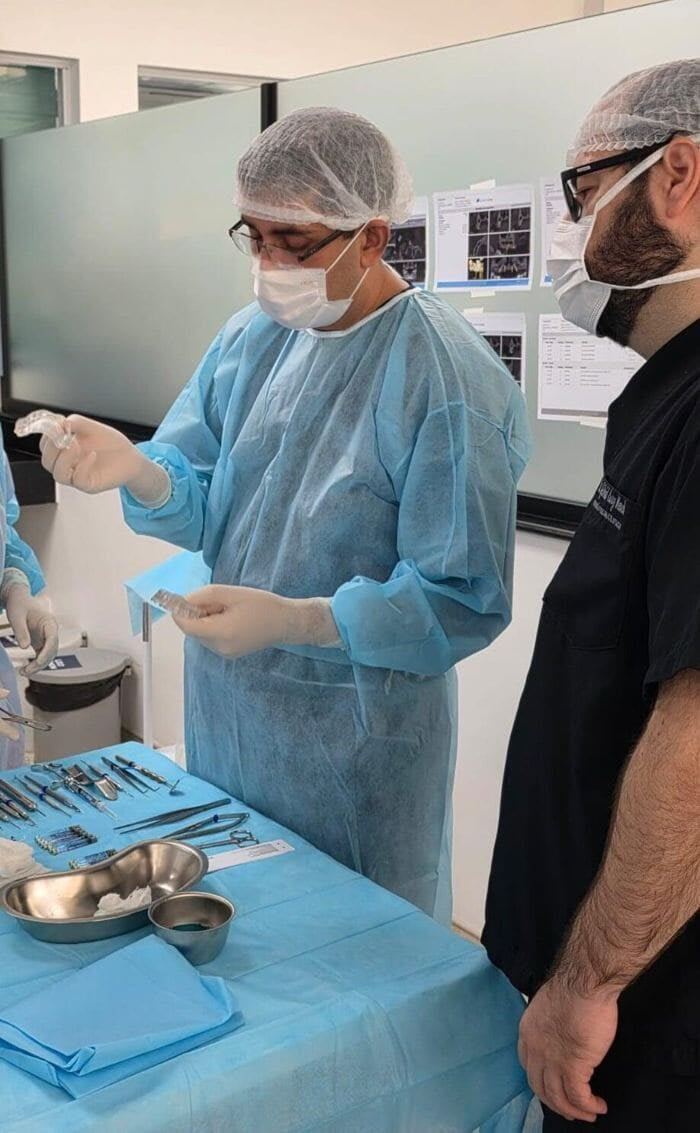
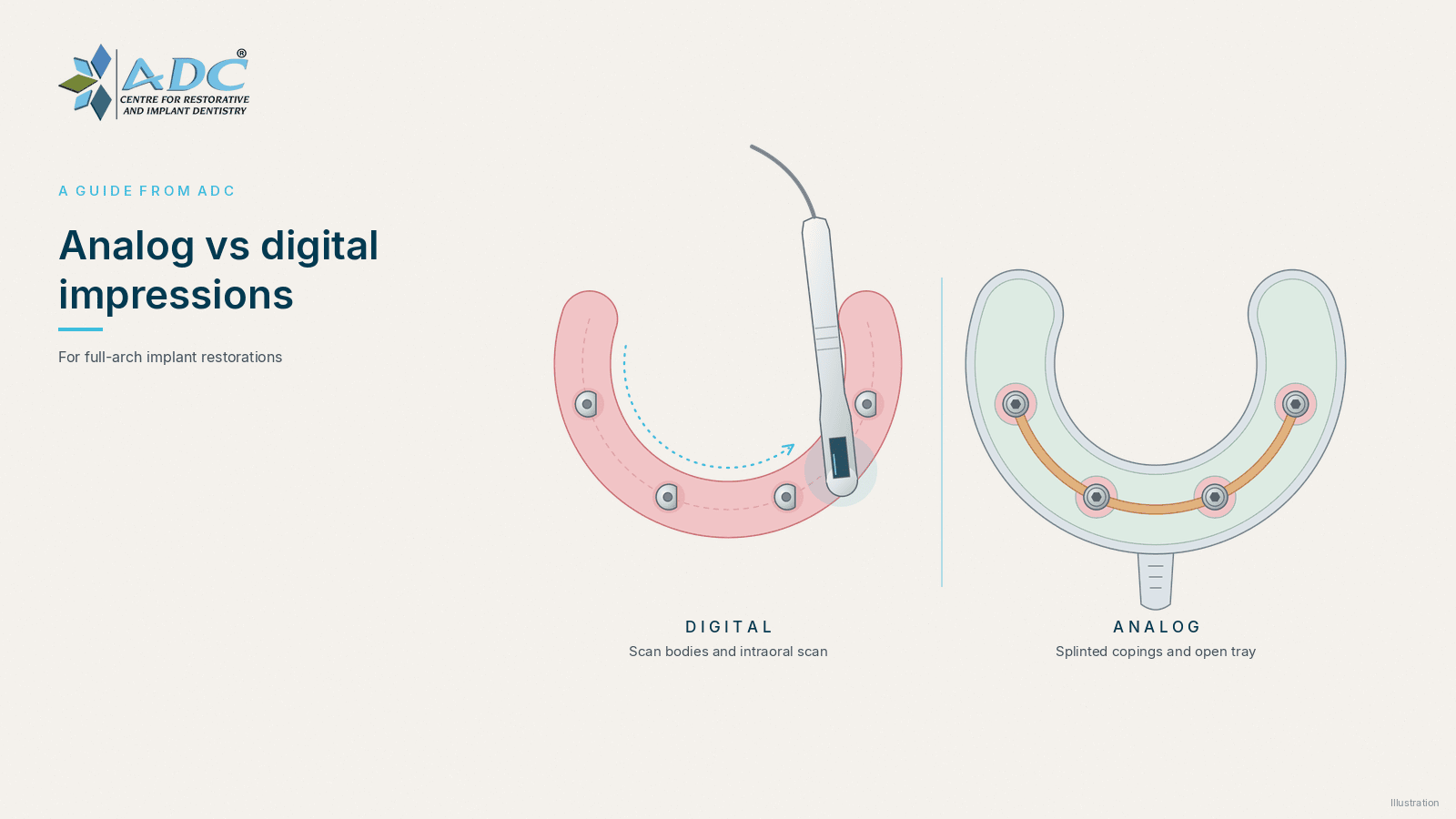
In the conventional workflow, impression copings are attached to the implants or to multi-unit abutments. For multiple implants, an open-tray (pick-up) technique is commonly used: the copings are screwed in place, the impression material is set around them, the coping screws are released through windows in the tray, and the copings come out inside the impression. Implant or abutment analogs are then attached and a cast is poured.
Practical points that affect accuracy:
- Splinting the copings. Joining the copings with a rigid material before the impression limits their movement. An in-vitro study on an edentulous model found less deviation with the splinted implant-level technique than with the non-splinted one.
- A rigid tray and a rigid material. A custom tray with enough space and a rigid elastomer such as polyether or addition silicone reduce distortion.
- Careful analog attachment. Rotating a coping while tightening the analog can move it inside the set material.
- Laboratory steps. Material shrinkage, tray flex and stone expansion all add small errors before the framework is made.
The analog route needs no scanner, but it has many steps where small errors can enter.
The digital workflow: scanner wand and scan bodies
In the digital workflow, scan bodies are screwed onto the implants or multi-unit abutments and captured with an intraoral scanner. Design software matches each scan body to its library file to locate the implant.
A systematic review on intraoral scan bodies describes them as playing an essential role in the digital workflow, and notes that design, fit and the scanning technique all matter. Points to check:
- The right scan body for the implant system, connection and abutment level, tightened as the manufacturer recommends.
- Complete seating, confirmed with a radiograph where the platform is subgingival.
- Condition of the scan body. Worn or damaged scan bodies should not be reused.
- A dry, clear field and a consistent scanning path, following the scanner manufacturer's guidance.
The edentulous arch is the hardest situation for intraoral scanning. Smooth, mobile mucosa offers the software few features to stitch images together, and the stitching error can grow over a long span. Some workflows add auxiliary reference features between the scan bodies or splint them to help the software. A digital record also has other parts: scans of the soft tissue, the opposing arch, the bite record and often the existing denture or interim prosthesis, so that tooth position and vertical dimension carry over into the design.
What the evidence says
A systematic review and meta-analysis comparing digital and conventional implant impressions reported that, for completely edentulous arches, digital scans appear to have accuracy comparable to conventional impressions. Its authors note that this conclusion rests mainly on in-vitro studies, and they recommend clinical trials before digital implant scans are adopted as routine for these cases. An earlier in-vitro study on an edentulous model reached a similar conclusion for accuracy. A separate systematic review of patient-centred outcomes looked at patient preference and working time for digital and conventional impressions.
Neither workflow removes the need for careful technique and verification.
Choosing a workflow for the case
When planning, consider:
- The number, depth and angulation of the implants, and whether multi-unit abutments are used.
- The state of the soft tissue and how much of the arch is featureless mucosa.
- The team's experience with each workflow, and how the laboratory receives and uses the data.
- Whether a combined workflow suits the case, for example a digital scan followed by an analog verification step, or a conventional impression digitised by the laboratory.
- How implant position will be verified before the definitive framework is made.
Verify before you finalise: a checklist
- Confirm seating of every coping or scan body, with radiographs where needed.
- Inspect the record. Look for voids, pulls or movement in an impression, and for gaps, distortions or stitching problems in a scan.
- Use a verification index or a try-in prosthesis. A rigid index or a printed or milled try-in should seat passively before the definitive framework is fabricated.
- Test for passive fit. Tighten one screw at a time and look for lifting at the other abutments, and check seating radiographically.
- Record the components used, the scan body or coping type, and any remake, in the clinical notes.
Frequently asked questions
Is a digital impression enough on its own for a full-arch implant case?
It can be part of a reliable workflow, but the evidence for full-arch scans comes mainly from laboratory studies. Many clinicians still verify the implant positions with an index or a try-in prosthesis before the definitive framework is made.
Open tray or closed tray for multiple implants?
For multiple implants, the open-tray technique with splinted copings is commonly used, because the copings stay fixed in the set material and are not repositioned by hand.
Do scan bodies need to be replaced?
Follow the manufacturer's instructions on reuse. A worn or damaged scan body can change the recorded implant position, so check its condition before each use.
Learning implant impressions with ADC
The ADC E-Learning Hub is the online training platform for dentists run by Dr. Mohit Dhawan and Dr. Rosy Dhawan. Its courses include Analog Impression Vs Digital Impressions in Implant Dentistry, the Basic Implantology Course, Hardware Complications in Dental Implants and a course on long-term implant success beyond osseointegration. Lessons are video-based with downloadable notes, a digital certificate is awarded for each completed lecture and module, and offline clinical classes are also offered, with pre-enrolment online. Browse the full list at All courses. For how misfit and load affect the screw joint, read Abutment screw loosening, and for choosing a course, read Implantology courses after BDS.
This article is general education for dental professionals and students. It is not individual clinical advice, and it does not replace the manufacturer's instructions, formal training or individual clinical judgement.


 International Team for Implantology
 ISO 9001:2015 Certified
 OHSAS Certified
 Academy of Osseo-Integration
 Indian Prosthodontics Society
 Indian Endodontic Society
 Women Implantology Network
 Indian Dental Association






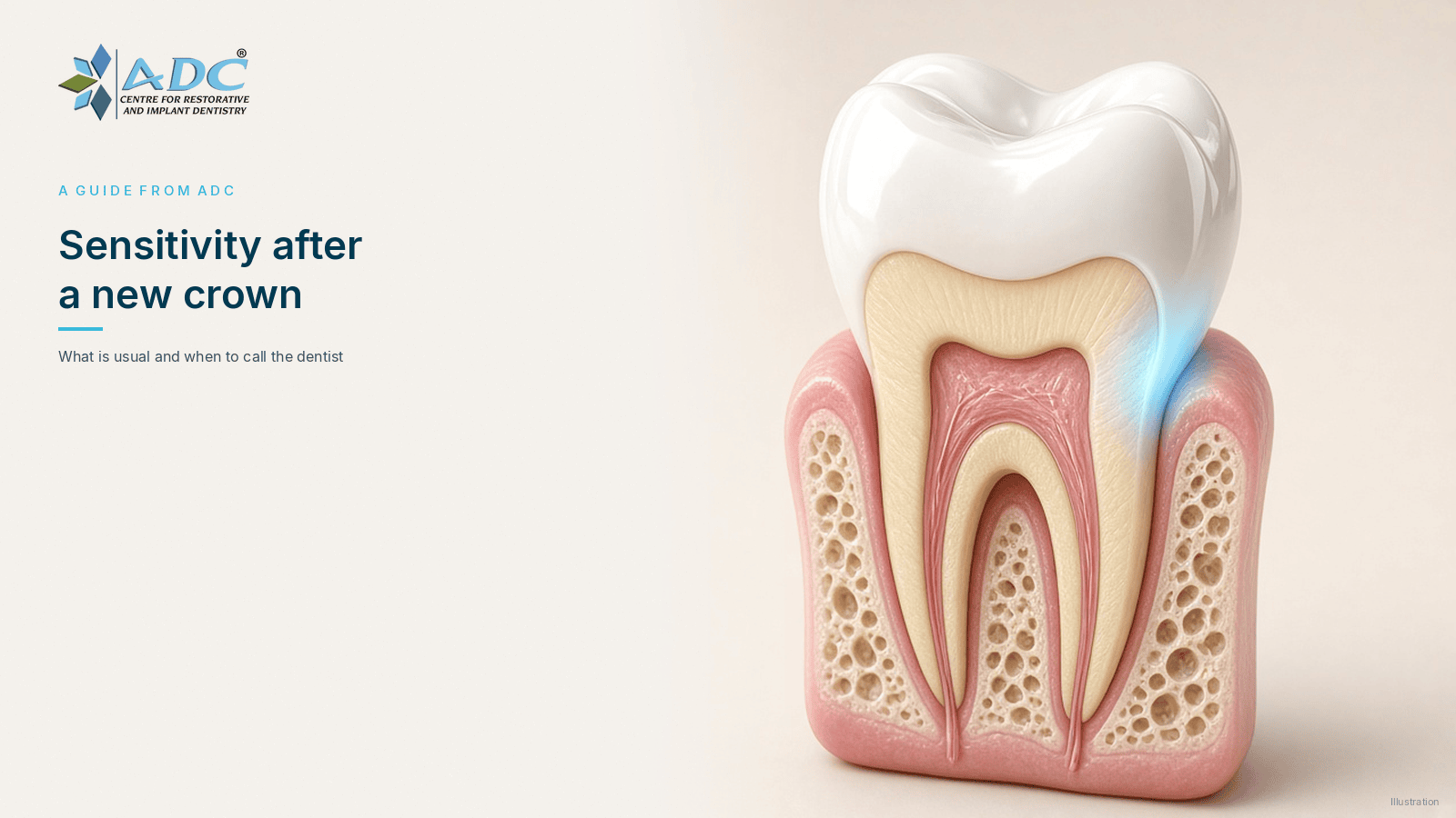 Sensitivity after a new crown: what is usual and when to call the dentist
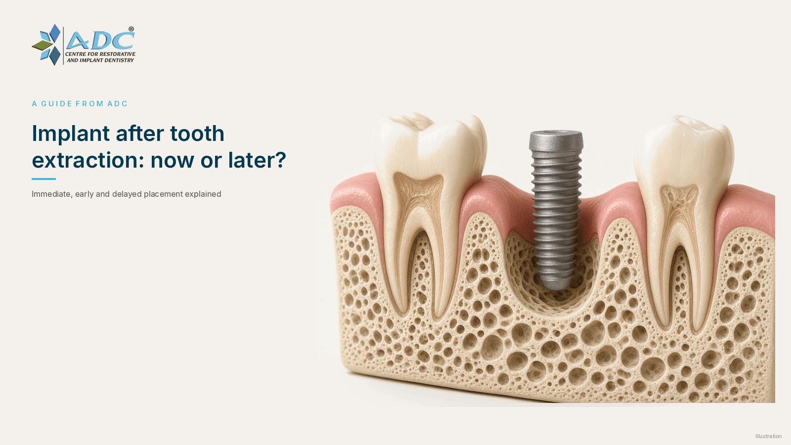 Dental implant after tooth extraction: immediate or delayed placement?
 Do you need a crown after root canal treatment?